Archive for the 'MRI' Category

2016 MRI

Posted by on Apr 11 2016 | MRI , MS

2016March12_T2AxialFlair Periventricular

19May2012

A couple of weeks ago I received a copy of the recent MRI images and report in the mail. There is lots of good news in it. No new lesions, no loss of brain volume. And the best news is that there are no longer any visible lesions in my cervical spine. Every previous report mentioned a lesion at C1. And there is no mention of edema in the spinal cord at C5/6 where I have a spinal stenosis.

Above I have posted images from this last MRI and the one in 2011 at the same level. It does look to me like the lesions are smaller.

Below are the images from the past 2 years: the one on the left 2016 and the one on the right from 2015.

2016March12_T2AxialFlair Periventricular

21March2015 periventricular T2 flair

These are encouraging results and I will discuss more with my neurologist at our upcoming appointment.

no comments for now

Latest MRI and neurology appointment

Posted by on Apr 16 2015 | Health Care , MRI , MS

21March2015 periventricular T2 flair

19May2012

Eight days ago, I received the MRI report and CD in the mail, and then the next day I had an appointment with my neurologist. It’s all good news as far as the MS goes. No new lesions, no change. And the neurological exam has even improved. So, officially this makes me NEDA or “no evidence of disease activity”.

I haven’t had a chance to really examine the images closely until today. The two images above are from as close to the identical slice through my head as I can get. The image on the left is the newest one (March 21, 2015) and the one on the right is from my very first MRI (May 19, 2012.) It’s encouraging to see that things look much the same. I am not sure if the greater darkness of the new image makes some of the lesions look smaller but it did give me some encouragement.

The neuroradiologist who helped me look more closely at the images has given me a couple of names of other neuroradiologists at VGH so I can view the images more closely and ask specific questions. I had requested detailed reporting on the most problematic lesions but the report only stated that there was no real change. And although that’s good news, I’m looking for visual evidence of healing!

no comments for now

Reading MRIs

Posted by on Nov 20 2013 | MRI , MS

Multiple sclerosis MRI Brain

Someone on one of my facebook groups posted this today and I thought it was an excellent ‘tutorial’ on reading MRIs and recognizing the patterns of all sorts of diseases and disorders.

It’s called Radiology Assistant and this is the direct link to the page on multiple sclerosis in neuroradiology . It’s published by the Radiological Society of the Netherlands.

Perhaps I should send this to neurologist #3 . . .

no comments for now

Nice diagram of the Vagus Nerve in medulla

Posted by on Nov 09 2013 | Health Care , MRI , MS

DorsalMotorVagusX

I pulled this diagram from Medical Neurosciences at the U of Wisconsin as it illustrates the complex and broad function of the vagus nerve (cranial nerve X or the tenth cranial nerve).

I had pain in my heart (called heartburn when in the hospital, although I did not agree with that description — I couldn’t argue with Doctor M), I had trouble swallowing (esophagus), I had trouble digesting fats (liver), I had food sit in my stomach for a long time (gastroparesis) that I would taste hours later, especially when sleeping. I lost my ability to defecate on my own (colon).

MS is a terrible disease. It seems to me that if one of my doctors had actually listened to me, they would have been able to diagnose the lesion affecting my vagus WITHOUT an MRI.

no comments for now

MRI review at VGH

Posted by on Nov 08 2013 | Health Care , MRI , MS

Two years ago, In addition to my facial pain, I had serious problems with my digestive tract — swallowing, regurgitation, constipation, gastroparesis, reflux. The weight loss led to hospitalization.

One year ago, I had a rapid exacerbation of my facial pain. The pain also led to hospitalization and where I finally got officially diagnosed with multiple sclerosis.

I wanted to see the MRI images in greater detail than I could see with the CDs I had ordered. So, yesterday, I met with the neuroradiologist at VGH who reviewed my first MRI from May 2012. At that time, he had been instructed to look for problems at the right side where the nerve root is. He didn’t see a problem there but reported on demyelination.

The images in the radiology viewing rooms are remarkable in their detail and clarity. I snapped a couple of shots with my camera so I could post them.

(If you’re really geeky — like me — and want to see what the real brain looks like in slices, there is a really nice website from UBC neuroanatomy with images taken at UVic .)

PontineLesions

Red – right-side lesions, blue – left-side lesion

This is an image of a slice through the pons. You can see that the right-side lesion (circled in red) is larger, as there are at least two lesions there. The one on the left side (circled in blue) may account for the facial numbness I experienced in 2005. I do have some sensitivity to cold in my upper left molars that could also be associated with it.

MedullaL

left-side lesion at medulla

This photo is a lower slice, through the medulla. The lesion there is large. This is where the vagus nerve originates and this would probably account for the gastrointestinal problems I had.

The neuroradiologist there is also a Dr. K and he was very nice. He invited me to contact him again if he could help.

I seem to have good experiences with doctors with K names!

Maybe I’ll call him Dr. K-3.

no comments for now

Neuroradiologist review of my MRIs

Posted by on Oct 21 2013 | Facial Pain/Trigeminal Neuralgia , Health Care , Letters to doctors , MRI , MS

neuroradiology

I recently asked an experienced neuroradiologist to review my MRI from 19 May 2012 as I wanted to know if my brainstem lesions were visible on that MRI. I wasn’t sure if I would even get a response. I am so amazed and impressed. I have encountered the extremes in our healthcare system but once in a while the response is more than you could hope for. He was generous and very kind in his willingness to help me out.

In the letter I had written to him:

I am requesting a favour from you . . . I would like to know two things.

• was the lesion in the pons visible on the first MRI?
• was the lesion in the medulla visible?

As these lesions would explain my facial pain and my GI problems, I wanted to fully understand the negligence of neurologist #3.

On Thursday, October 18, I received a response. He reviewed all three of my scans:

In answer to your question, both the lesion(s) on the right side of the pons (there is probably more than one) and the lesion in the medulla were visible on the first MRI, although not as clearly shown as on the MRI at UBC which was performed with a dedicated MRI protocol and somewhat thinner slices.

Your case is unusual although certainly not unheard of, in that you have quite a number of lesions on your brain MRI (many of which appear old) with little in the way of “classical” symptoms and signs.

That tells me a lot. Yes, my MS is old. And yes, my presentation is unusual. Neurologist #3 had everything he needed after that first MRI to diagnose my MS and my pain. Perhaps neuroradiologists should be given the authority to diagnose MS and remove it from the hands of neurologists? I am sure that we would get earlier diagnoses that way. In my case, it would have saved the system a lot of money.

And at least two lesions on the right side of the pons — yikes. No wonder I had so much pain.

1 comment for now

Do I have CCSVI?

Posted by on Oct 07 2013 | CCSVI/Neurovascular health , Facial Pain/Trigeminal Neuralgia , MRI , MS

After meeting Lori Batchelor last month and thinking about the logic of having good blood flow for a healthy brain, I decided to go to AIM Medical Imaging to see whether or not I had narrowed veins. I had heard that Dr. Raj Attariwala was the best diagnostician in Canada, so I booked an appointment for September 30.

The McMaster Study showed only one person with MS who had CCSVI out of a hundred. I was eager to challenge that finding as I suspect that many people who do not have MS also have CCSVI. And if it’s only 1/100 for people with MS, then chances were slim that I would have it. It was interesting to note that they were looking specifically for vein blockages, not reduced flow or backwards flow/reflux.

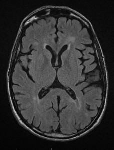
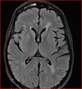
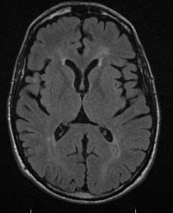
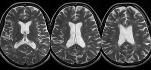
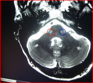
Immediately after the scan, I had a consultation with the radiologist, Dr. Attariwala. I had given him copies of my previous MRIs to look at. Fortunately, there were no new lesions or active lesions. 
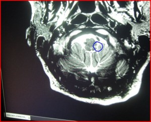
He was able to identify the lesion in my pons from the very first MRI in May of 2012. (I wish I had paid for this MRI two years ago as it would have helped guide me better than all the specialists I saw and tests I had. It would have been faster and cheaper and I probably would not have ended up hospitalized twice.)

Within a couple of hours, I had images and results. My right jugular vein flows significantly less than my left. This is the opposite of the findings at McMaster where everybody had greater flow in the right than the left. He says that this is a finding that has been “known for years.”  (from extended interview with Dr. Rodger ) Jugulars

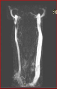
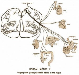
The central vein is called the ‘azygos’ — it was described to me as ‘awful’.

So, it appears to me that I do have CCSVI or chronic cerebrovascular insufficiency according to these images and flow measurements. However, I am not knowledgeable enough to know or assess if I meet the full criteria for CCSVI. This is how it was described in the report:

INDICATION: History MS, query jugular venous narrowing.

TECHNICAL Conventional study: axial flair, T2, 3-D space flair, post gad T1sagittal T1 , PD, axial T2 C-spine.

Research: SWI, Venc, azygous, with subtracted multi-sequential venogram through the neck and lower head,

FINDINGS: The patient recognizes that jugular MRI venogram as well as iron deposition sequences remain in the realm of research . . . Known MS demyelination, without MRI evidence of active lesions. To be determined by ongoing research; there is associated substantia nigra iron deposition which is of uncertain significance. The jugular veins demonstrate asymmetry of flow, with the right being diminutive, with transient halting of flow, without reversal. These findings plus the controversial nature of the research component of the study was discussed with the patient.

In the images, I received a graphic representation of the flow measurements taken.

The red line represents flow in the right, the green line in the left. If the red line were to go above the horizontal line at 0, it would indicate backflow or reflux. So, the next step is to investigate further.

There are so many unanswered questions about the procedure and I’m not convinced that some of the research currently being conducted is going to lead to good answers. Most of the research has been looking for gross measures of correlation between blood flow and ways to measure it and assess it, then make pronouncements about the lack of connection with MS, end of story!! For people with MS who have benefited and thrived after the procedure, it has clouded the debate. So many people are looking for a way to end this area of research, calling it a social media phenomenon of unsubstantiated anecdotes. But they are choosing to ignore evidence right in front of them.

I think we need to ask better questions. What’s happening when people get good results from venoplasty? Why aren’t we studying them? Why is there no single-case research being done on an amazing case like Lori Batchelor? Her neurologist is on camera saying that there is improvement when she was diagnosed with secondary progressive MS. How does that happen?

no comments for now

One-year anniversary of knowing for sure

Posted by on Aug 10 2013 | Facial Pain/Trigeminal Neuralgia , Health Care , MRI , MS

Last year, on Friday, August 10, at 11:36 am, I received the written MRI report via e-mail from neurologist #3’s assistant. When I read the report, it was 100% confirmed for me that I had MS. Although they had used the term ‘demyelination’ in the previous MRI, I still knew what that meant. But there is something about seeing the words ‘multiple sclerosis’ in the radiology report that makes you realize that denial is no longer an option. While it described the lesion in the left medulla that would be connected with my GI issues (affecting the vagus nerve), it did not describe a lesion in my right pons. I had hoped for that so that there would be concrete evidence for the source of my facial pain. The radiologists did their job as it was a routine bran scan — they alerted the neurologist #3 to what they were seeing and it was his responsibility to look at it more closely. (I needed to wait until November to have my MS neurologist look at this very scan closely to identify the lesion in the pons. I still can’t identify it on this scan on my home computer but I did find the lesion on the MRI I had earlier this year.)

received by e-mail 10 Aug 2012; click to enlarge

At this same time, Daniel was over at the office of the GP who had laughed at me to inform him that he would no longer be seeing him. (I had insisted on this because I did not want my husband to be ‘cared for’ by a doctor who could treat anybody the way he treated me.) So, when he came home, I had something to discuss with him. But to my surprise, the GP #3 told Daniel at that meeting that I should stay in touch with my ‘psychologist friend’ as it looked like I had MS. (Even though I had communicated my GP #4 change in January, this previous GP #3 continued getting reports and he felt at liberty to talk to my husband about it — astonishingly unprofessional! Graduate of UBC Medical School, 1985)

I contacted GP #4 right away and made an appointment to get a referral to the MS Clinic. I had already given up on neurologist #3 as he had done nothing to help me figure things out, but I still knew it was his responsibility to review the MRI with me since he had ordered it. And since it indicated MS, I believed that I would be able to get an appointment within a couple of weeks. But I requested an appointment on Monday the 13th, and was given an appointment for two months later, on October 9.

I moved into action. I reviewed the neurologists at the MS Clinic and chose one to be referred to and prepared the referral information for my GP so that it would be an easy referral for her to make. That was completed by August 14. I looked for information on MS support groups and joined one that meets weekly at Kitsilano Community Centre on August 24. I took advantage of the workshops offered by the MS Society. I met and talked with friends of friends who had MS to gather information.

The MS Clinic appointment was originally scheduled for December 13, 2012. I stayed at home and waited, armed with drugs for facial pain. Then I did what I could with dentists and oral medicine specialists over the next couple of months to rule out other problems with my dental pain, until we reached the conclusion in October that the pain was most likely from the MS. I still wanted the concrete evidence.

On Friday, November 16 2013, when neurologist #5 came into my room at UBC hospital as I lay there in constant pain/misery, to let me know that he had identified a lesion in the right pons. I got up, walked over to the door, shook his hand to thank him, then crawled back to bed.

I had the answer I had been looking for.

no comments for now

Dr. Chris Honey on trigeminal neuralgia and multiple sclerosis

Posted by on Aug 01 2013 | Facial Pain/Trigeminal Neuralgia , MRI , MS

I was thinking about my interactions with our local neurosurgeon, who has remained unnamed in my blog so far. I did refer to him in a post after receiving my medical records, as well as my disappointment with his grand rounds at St. Paul’s hospital after I had talked to him on the phone (while in pain) about what I hoped he would say to the young medical practitioners. A good friend of mine suggested that he is a possible candidate for burnout considering the complexity of his professional life. It’s important for me to remember that doctors are human too. Dr. Honey is smarter than I could ever hope to be, unbelievably accomplished and generous with his time and knowledge as well.

I visited Dr. Honey’s website again today and was pleased to see that there is additional information on it. He gives some good preliminary information on trigeminal neuralgia in multiple sclerosis and more detailed information on his page on trigeminal neuralgia . The info is solid, thorough and current.

It has been reported in the literature that people have starved to death from the pain of TN. On his website, Dr. Honey talks about Dr. Johann Bausch, a doctor who starved to death from TN . I believe he’s referring to me in this comment:

Readers will be horrified to know that one of my patients reported that she almost starved to death before the diagnosis was made and she received treatment in 2012. History repeats!

2014 Update **VIDEO NO LONGER AVAILABLE** accidentally deleted by Adobe

I was delighted to see that the Trigeminal Neuralgia Association of Canada has given Dr. Honey the link to the excellent webinar he gave on March 30, 2013. I had the chance to ask a question at the end at 58:00 where I asked “What do you do if your neurologist doesn’t look at your MRI?” and he responded, “get another neurologist”, which is what I ended up having to do. He talked about some neurologists being “a little old school” but it seems to me that there are continuing education requirements for all professions. It’s heartbreaking to learn that we have highly-paid professionals who neglect that responsibility. For those of us out there suffering because of that — thank god for the internet!

Thanks Dr. Honey and TNAC for making this wonderful webinar available.

1 comment for now

Medications and MRI

Posted by on Jun 04 2013 | Health Care , MRI , MS

windows at the new Brain Research Centre at UBC

Daina and I went to UBC this morning for a session on medications for MS provided by one of the nurses at the MS Clinic. It provided me with some answers to questions but ultimately the choice is mine with what I choose or whether or not I go on the drugs. They like to say that MS is ‘manageable’ now and that people can live long and full lives with it. The key is to find what works to stop the progression of the disease. Ideally, I should have been in this position several years earlier . . .

Later in the afternoon I went to meet with the MS neurologist. While I was in the waiting room, Dr. Perry came out to the reception area with a medical student. I said ‘hi’ and the next thing I knew, he was inviting me to an event on Thursday evening for the Therapeutics Initiative program.

Neurologist #5, my MS neurologist, reviewed my MRI in detail with me. He identified the lesions, described several of the older lesions that were ‘cavitated’, and pointed out the lesion in the pons that caused my trigeminal pain. I blogged about the MRI in a previous post and had correctly identified the location of that lesion.

lesion in pons

We also took a look at the major arteries in my brain and he described them as ‘pristine’ with no signs of arteriosclerosis or malformations. So, my risk for stroke is minimal. Kind of ironic as I don’t want to live a long life any more. The major ‘take-home message’ for me was the fact that my MS lesions don’t show evidence of healing, as some other people’s do. So, my best defence is to prevent new lesions. That will be an important factor in the choice of medications.

no comments for now

Next »