MRI from July 2012
Tomorrow I will be going into my GP’s office to get the results of my recent full MRI. They received it before I did. I’m not sure if they get the images or just the radiology report. It does assure me that my copy will be in the mail shortly.
I spent some time reviewing my MRI images from July. These are the images that the ordering neurologist (neurologist #3) didn’t bother to look at. Having studied the brain and worked with people who have had strokes and brain injuries, I was able to identify some of the classic lesions seen in MS easily.

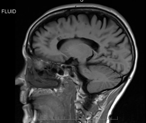
Sagittal view, featuring lesions in corpus callosum
Lesions in the corpus callosum are one of the classic signs of MS. The corpus callosum is a very thick band of myelinated nerve fibers that connects both hemispheres of the brain. You can see the slice through the corpus callosum above the black arc (lateral ventricle) in the middle of my head. The imaging technique used for this view shows lesions as dark spots.

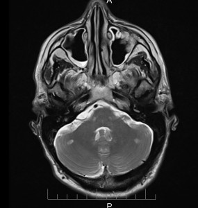
lesions (white spots) in cerebellum
This picture is an axial view, meaning that it’s a slice of my brain from front to back, with the nose on top and back of my head at the bottom. Lesions in the cerebellum are also classic in MS. The cerebellum is the lower part of the brain behind the brainstem and is responsible for coordination of movement. The imaging technique used for this image shows the lesions as white spots.

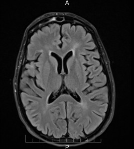
periventricular lesions
This image is the same orientation as the one before but higher up in my brain. Again, the lesions are white. Lesions around the ventricles (large black curved spaces) are classic in MS. The ventricles are connected holes in the brain that contain cerebrospinal fluid.
I don’t know if you can imagine how hard it was for me to sit in neurologist #3’s office on October 9, 2012, as he told me that I didn’t have MS and that constipation has nothing to do with MS. I had looked at the images and he hadn’t. It is the height of arrogance to assume that the radiologists he works with don’t know what they’re talking about and not bother to look himself. Interesting to note how he can say one thing to my face “you don’t have MS” and then write a report that states he has actually considered what the radiology report says.
“Her MR brain scan completed in July 2012 was reported to be consistent with a diagnosis of multiple sclerosis. . . The findings from the scan are surprising. She has never had any neurological deficit on examination and her presentation is most unusual.”
I guess if you only have deficits that are sensory (numbness/pain) or invisible (gastrointestinal), you need to have a neurologist who listens and believes you. I can only assume that he did not believe a word I said, probably even more likely after the reports from the hospital stay in December 2011/January 2012 came out with the gastroenterologist who made all the errors in my intake and said I had post-herpetic neuralgia (nerve pain after a case of shingles when I have never had shingles) and a conversion disorder.
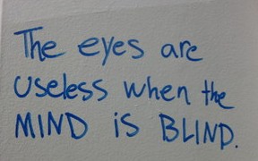
As the graffiti in the Granville Skytrain station stated:
I haven’t written about the hospital stay in detail yet. I guess I need to.
3 Responses to “MRI from July 2012”
Trackback URI | Comments RSS

[…] are the brain images of the last MRI on March 4, 2013. I chose images similar in orientation to the ones from July that I wrote about here and that show the lesions well. Sagittal view, T2 […]
02 Apr 2013 at 5:16 pm
I truly believe that radiologists look at so many films during the day that they just scan them way to quickly and unless the lesions are in their face they tend to overlook them. I have copies of my MRI’s where they see no enhanced lesions, when you take time to look you can see them, mine just don’t show in your face, as I’m sure a lot don’t, especially spinal lesions which are hard to see. You need to study online lesions if you think you have MS and take time to check yourself. DON’T read into the films. Education with this disease is the key.
I am a retired nurse and truly feel that you know your body, and God has a way of telling you what is wrong, you just have to listen.
13 Jun 2014 at 10:46 am
I agree Karen, some of the smaller ones might get by them but classic MS lesions in the brain were observable in my case. They reported on them. It was the neurologist who bungled my case and delayed the diagnosis by not investigating further.
13 Jun 2014 at 11:00 am